
The functionally most important parts of the penis are the corpus cavernosum and the corpus spongiosum. The corpus cavernosum arises with paired cavernous bodies from the inferior pubic branches (Fig. 1.1). In front of the symphysis, the two corpora cavernosa unite to form the corpus. In humans, with few exceptions of cavernous body separation [3], there is an incomplete cavernous body septum, which allows blood transfer between the corpora and thus makes the cavernous body a functional unit.
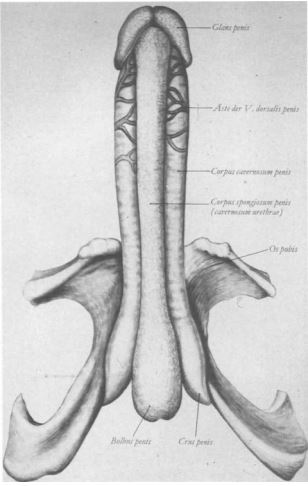
Fig. 1.1. Corpus cavernosum of the penis in erect state (from [1]).
A very firm connective tissue sheath, tunica albuginea, about 1 mm thick, surrounds the cavernous tissue. The spongy tissue of the corpus cavernosum consists of a network of connective tissue bars containing smooth muscle, covered on the surface by endothelium, and delimiting a labyrinth of fine venous blood spaces called lacunae [1]. Tunica albuginea and the connective tissue of the corpus cavernosum form the fibrous skeleton of the corpus cavernosum which serves for stabilization [4].

Fig. 1.2. Transverse section through the male penis.
The blood supply (Fig. 1.2) is arterial, mainly via the profunda penis artery, a terminal branch of the internal pudendal artery (from the internal iliac artery), which lies inside the corpus cavernosum. Branches from the dorsalis penis artery serve to supply the glans, but may also contribute to the supply of the corpora cavernosa through perforating branches [2]. Venous outflow is predominantly via the dorsalis penis profunda vein and via crural veins localized in the depth of the corpora cavernosa, which drain to the periprostatic plexus.